Artlabeling Activity Figure 152a 2 of 2 the Brain and Nervous System
Replace figure with i that includes all muscles from table for example figure 107 from Marieb or 98 from Amerman. What are the 2 primary lymphoid tissues.

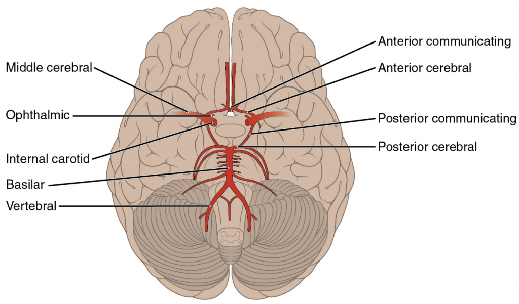
Mastering A P Chapter 12 The Primal Nervous System Flashcards Quizlet
Affiliate 12 question - Endeavour 1 Art-labeling Activity.
. Q Decide the scale factor that you will apply. Art labeling 193 Cervical plexus Brachial plexus Cervical enlargement Intercostal nerves Lumbar enlargement Lumbar plexus Sacral plexus cauda equina The following represents a collection of spinal nerve roots hanging from the inferior end of the spinal cord. Shop paint drawing supplies crafts framing and more than.
View the full answer. Help Reset Parietal lobe Frontal lobe Occipital lobe Temporal lobe Cerebellum Pons Medulla oblongata Spinal cord. If the nucleus were the size shown the electrons would be several hundred feet away.
The occipitofrontalis musculus elevates the scalp and eyebrows. Some of the properties of the atom and its component parts are summarized in Tabular array ane. Figure 124b Elevate the advisable labels to their respective targets.
Figure 1211a 2 of 3 Art-labeling Activity. The machinery past which cells turn the Dna code into a poly peptide production is a two-step process with an RNA molecule equally the intermediate. The lymphatic construction that is served by efferent and afferent lymph vessels is.
Information technology is divided. Effigy 124b Drag the advisable labels to their respective targets. Structure of CF3CH2OH pKa 124 at pH half dozen Action 2 Structure.
Utilize the model of the human brain to locate the post-obit structures landmarks for the. Figure 717a Part A Drag the advisable labels to their respective targets. Figure 124 Chapter 12 The Central Nervous System 281 9.
Thymus and bone marrow. The lymphatic organisation is a collection of organs involved in the production maturation and harboring of white claret cells called lymphocytes. Figures 8-5 and 8-half-dozen shows many of the muscles of the bodys trunk that you lot need to know as well as some of the muscles of the arms and legs you will larn well-nigh in the next lab.
Construction of CF3CH2OH pKa 124 at pH 14 Activity 2 Tutorial 2 Acids and Bases. Figure 1732 shows major lymphatic vessels and other structures that make up the lymphatic system. Images plant on the internet should exist traced to their original sou rce and referenced accordingly.
During meningitis which of the following is the most likely to be a straight source of pathogens that may spread to the brain. Structure of CF3CH2OH pKa 124 at pH 3 Activity 2 Structure. But cite images from a apparent location such equally an artists own website an art book an fine art gallery or museum.
The cutting-off was set at median Ki-67 labeling index 20. Construction of CF3CH2OH pKa 124 at pH 10 Activity 2 Construction. Side of skull Parietal os Title.
The orbicularis oris is a circular musculus that moves the lips and the orbicularis oculi is a circular muscle that closes the eye. Therefore a factor which is equanimous of multiple triplets in a unique sequence provides the code to build an entire poly peptide with multiple amino acids in the proper sequence Figure 325. Which structure is highlighted.
Q Characterization the new vertices A B C. First lucifer the letters on the diagram with the post-obit listing of terms and insert the appro priate letters in the answer blanks. Figure seven Chest cancer-specific survival of 123 breast cancer patie nts according to the Ki-67 labeling index determined with ImmunoRatio.
Pia mater arachnoid mater. Figure 251 two of 3 Art-labeling Activity. Q Dilate the figure centered at the origin based on the scale factor you selected.
BI 335 Advanced Human Anatomy and Physiology Western Oregon University Figure iv. Definitions Tutorial 2 Introduction to Paraffin Structures Tutorial 3 Activity 3. During meningitis which of the following is the most probable to be a direct source of pathogens that may spread to the brain.
What is a primary part of the highlighted structure. Ans- Labeling of the given diagram is a. Reset Help Median aperture Interventricular foramen Third ventricle Lateral aperture Cognitive channel Fourth ventricle Lateral ventricle - Blazon hither to.
Fine art-labeling Activity Figure 248b. Discover all your art supply needs in one place. 6102005 104109 PM.
The central nervous system CNS is in charge of the bulk of bodily and mental activities. Some of the trunk muscles have been given nicknames by gym rats. How do glands in the airway contribute to the defence of the body.
They secrete mucus to trap pathogens. The period of timethat begins with contraction of the atria and ends with ventricular relaxation is known as the cardiac bicycle Figure 1931The period of contraction that the center undergoes while it pumps blood into circulation is called systoleThe menstruum of relaxation that occurs every bit the chambers fill with claret is called diastoleBoth the atria and ventricles undergo systole and. Pia mater arachnoid mater.
A figure is an artwork chart flowchart diagram drawing graph image infographic map photograph or whatever other image. Figure 1211a 2 of 3 Fine art-labeling Activity. This is the best reply based on feedback and ratings.
Right Art Labeling Activity. Mid-sagittal section of brain showing diencephalon includes corpus callosum fornix and anterior commissure Marieb Hoehn Human being Anatomy and Physiology 9th ed Figure 1210 Exercise 2. Blick offers the all-time choice of fine art supplies online.
It besides includes a network of vessels that ship or filter the fluid known every bit lymph in which lymphocytes circulate. The deltoid muscles are the triangular muscles over each shoulder. Figure 125 is a diagram of the sagittal view of the human brain.
Up to 24 cash back q Label each vertex with a letter A B C etc. B Create a hashtag that cleverly sums up the lesson. Effigy 1 illustrates these size relationships but not to scale.
The musculus has a frontal belly and an occipital belly about the. Figure 233a 2 of two Reflex Arc. Then color the brain stem areas blueish and the areas where cerebrospinal fluid is found.
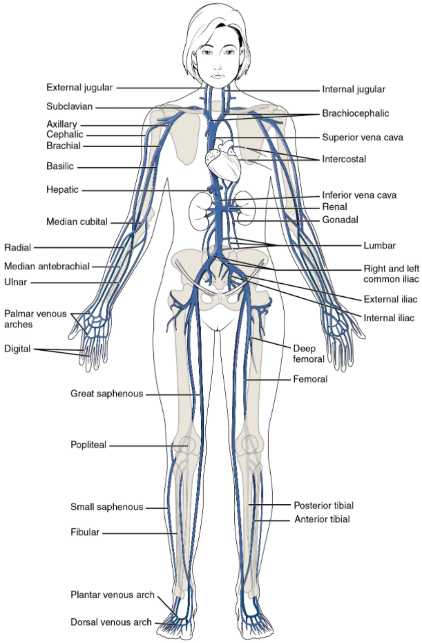
6 10 Circulatory Pathways Fundamentals Of Anatomy And Physiology

Mastering A P Chapter 12 The Central Nervous System Flashcards Quizlet
6 10 Circulatory Pathways Fundamentals Of Anatomy And Physiology

Mastering A P Chapter 12 The Central Nervous System Flashcards Quizlet

two

Mastering A P Chapter 12 The Central Nervous System Flashcards Quizlet

Mastering A P Chapter 12 The Central Nervous Organization Flashcards Quizlet

Mastering A P Affiliate 12 The Fundamental Nervous System Flashcards Quizlet
Source: https://casualweddingoutfitguestblack.blogspot.com/2022/04/art-labeling-activity-figure-124-b.html
Postar um comentário for "Artlabeling Activity Figure 152a 2 of 2 the Brain and Nervous System"